Created by Itzhak Brook MD a physician and a laryngectomee. It contains information about head and neck cancer, life after laryngectomy, and manuscripts and videos about Dr. Brook's personal experiences as a patient with throat cancer. It has information about side effects of radiation and chemotherapy; methods of speaking; airway, mucus, stoma, voice prosthesis; eating and swallowing; medical, dental and psychological issues; respiration; anesthesia; travelling; and COVID-19.
"My Voice"
Order a paperback or Kindle Edition or e-book of "My Voice: A Physician's Personal Experience with Throat Cancer," the complete 282 page story of Dr. Brook's diagnosis, treatment, and recovery from throat cancer.
Order a paperback or Kindle Edition or e-book of "The Laryngectomee Guide," the 170 page practical guide for laryngectomees.
Eating, swallowing, and smelling are not
the same after laryngectomy (also called laryngectomie, laryngektomie, laringectomia, laryngektomii, laringektomija, laringektomiya, and larenjektomi). This is because radiation and surgery create permanent
lifelong changes. Radiation therapy can cause fibrosis of the muscles of mastication which can lead to one's inability to open the mouth (trismus or lockjaw), making eating more difficult.
During
laryngectomy, certain structures in the throat important in the natural act of
swallowing are removed. Physically, swallowing is very different, since
reconstruction can limit movement of the tongue base, important in driving food
downward towards the esophagus. . It can also cause frequent biting of the tongue. Additionally, with the removal of the vocal cords
and the diverting of the trachea, subglottic pressure to drive food down the
esophagus no longer exists, so the throat muscles have to handle more of the
work.
Eating and swallowing difficulties can also be generated by a decrease in the production of saliva, and a narrowing of the neopharynx (new pharynx) and esophagus, plus the lack of peristalsis in those with flap reconstruction. Swallowing difficulties and painful swallowing can lead to accumulation of saliva and oral secretion in the mouth. Smelling is affected because inhaled air bypasses the
nose. Treatment of eating and swallowing difficulties following laryngectomy depends
upon the underlying cause.When it is
the result of reduced pharyngeal driving forces, the speech and language pathologist (SLP) can provide exercises
to strengthen the base of tongue.Additionally, patients can be provided with strategies such as soft food
preparation and liquid wash to facilitate pharyngeal clearance.In cases where the upper esophageal sphincter
does not open adequately or there is stricture, surgical procedures can reduce
obstruction.These procedures include surgical
division of muscle fibers (myotomy), laser resection of the scar bands, dilation
(surgical stretching), or Botox (surgical denervation).
This section describes the manifestations
and treatment of the eating and smelling challenges faced by laryngectomees. These
include swallowing problem, food reflux, esophageal strictures, and smelling
difficulties.
Maintaining
adequate nutrition and liquid consumption in a laryngectomee
Eating may be a lifelong challenge
for laryngectomees. This is because of swallowing difficulties, decreased production of saliva (which lubricates food and eases mastication), and an alteration in one's ability to smell.
The need to consume large quantities
of fluid while eating can make it difficult to ingest large meals. This is because
when liquids fill the stomach there is little room left for food. Because liquids are absorbed within a relatively short period of time, laryngectomees end up
having multiple small meals rather than fewer large ones. The consumption of large quantities of liquid makes them urinate very frequently throughout
the day and night. This can interfere with one's sleep pattern and can cause tiredness and irritability. Those who suffer from heart problems (e.g., congestive heart failure) may experience problems due
to overloading their bodies with excess fluid. Consuming food that stays longer in the stomach (e.g., proteins
such as white cheese, meat, nuts) can reduce the number of daily meals, thus reducing
the need to drink liquids.
It is important learn how to eat without ingestingexcessive amounts of liquid. Relieving swallowing difficulties can reduce
the need to consume fluids, while consuming less liquids prior to bedtime can
improve sleeping pattern.
Nutrition can be improved by:
•Ingesting adequate
but not too much liquid
•Drinking less liquid in the evening
•Consuming “healthy” food
•Consuming a low carbohydrate and high protein diet (high sugar
enhances yeast colonization)
•Requesting dietitian assistance Enclosed is a video that how to eat when one has swallowing difficulties:
It
is essential to make sure a laryngectomee follows an adequate and balanced
nutrition plan that contains the correct ingredients, despite difficulties with
their eating. A low carbohydrate and high protein diet that
includes vitamins and minerals supplements is important. The assistance
of a nutritionist, speech and language pathologist (SLP), and physicians in ensuring
that one maintains adequate weight is very helpful.
Fluid
consumption by laryngectomees
Fluid
consumption by laryngectomees is a balancing act between the need to drink them
and their potential adverse effects. Laryngectomy can lose fluids through
exhalation when they do not wear an HME.
Laryngectomee
require to drink fluids for these reasons:
Enhancing
swallowing
Maintaining moisture in the mouth to compensate for not having enough saliva (xerostomia)
Maintaining
adequate respiratory tract moisture especially in low humidity
Maintaining adequate fluid volume in their body (preventing dehydration, and orthostatic
hypotension)
Allowing adequate perspiration (especially in hot weather)
Enhancing urine excretion
Adverse
effects of consuming excessive amounts of fluids:
Frequent
urination that can interfere with sleeping (especially in men with enlarged prostate)
Overloading the cardiovascular system
Reducing
the amount of sodium in the blood (hyponatremia)
The
recommend daily consumption of fluids for adults is about eight 8-ounce
glasses, which equals about 2 liters, or half a gallon. However, this may vary
depending on environmental conditions (i.e., hot weather, low humidity), individual’s
needs and medications.
Excessive
salivation (sialorrhea)
Excessive salivary flow or sialorrhea can occur in patient
following oral or head and neck surgery. In this setting the sialorrhea may be
a result of over secretion (primary sialorrhea) of the salivary glands but is
more commonly due to impaired neuromuscular control with dysfunctional
voluntary oral motor activity that leads to an overflow of saliva from the
mouth (secondary sialorrhea). Patients often have inefficient and infrequent swallowing
(often due to pain or swelling), which further compounds the problem. It can
also be caused by tumor or treatment-related difficulties in swallowing (dysphagia),
or due to altered anatomy from oncologic resections of the upper aerodigestive
tract, especially the middle third of the mandible. Post-operative sialoceles
and fistulas are other manifestations of abnormal salivary flow which interfere
with wound healing.
Sialorrhea poses several risks to the patient. Excess saliva spilling over the bottom lip can
leading to drooling, poor post-surgical wound repair, external skin irritations
and breakdown, rashes, poor quality of life, and social ramifications. Potentially
more dangerous sequelae are those affecting the respiratory system. With an
inability to self-manage secretions, especially in patients with poor airway
control and clearance, the one or more liters of saliva produced regularly each
day can result in aspiration, choking, poor oxygenation, and life-threatening
pneumonias.
I most individual post sialorrhea is self-limited and can be managed
conservatively by suctioning or swallowing the secretions, reducing the post-surgical
pain that may interfere with swallowing and by sitting up allowing the
secretion to drool rather than be swallowed.
Working with a
speech and swallowing therapist can help manage the mechanics of swallowing and
reduce saliva accumulation. Maintaining good oral hygiene can help manage
saliva and prevent infections that may contribute to hypersalivation.
Various pharmacologic agents can be used in the management of
sialorrhea. None of these agents are specifically FDA-approved for sialorrhea
and are often used off-label. These include glycopyrrolate, scopolamine, atropine,
and benztropine. Botulinum toxin injection into the salivary glands has also
been used. In some cases surgery and radiation therapy are used to correct persistent
excessive salivation.
Belching (burping), hiccup, air trapping and flatulencein laryngectomees
Laryngectomees are prone to develop burping, hiccup and farting especially in the immediate period after surgery. One cause for this
is that the post-surgical narrowing of their upper esophagus may interfere with
the upward passage of swallowed air which is collected in the esophagus and stomach.
These issues may persist in many individuals.
Air trapping below the esophageal
narrowing is common in those who use voice prosthesis where some of the air directed
to the esophagus can collect. Also rapid swallowing of food and liquids (gulping)
can allow excessive air to collect in the esophagus and stomach. One may also swallow
excess air when eating or drinking too fast, talking while eating, chewing gum,
sucking on hard candies, drinking carbonated beverages, or smoking.
Acid reflux or gastroesophageal reflux
disease (GERD) can sometimes cause excessive belching by promoting increased
swallowing. Chronic belching may be caused by inflammation of the stomach
lining (gastritis) or an infection with Helicobacter pylori, the bacteria
that can cause stomach ulcers. In these cases, the belching is accompanied by
other symptoms, such as heartburn or abdominal pain. Poorly fitting dentures
can cause excess air swallowing when eating and drinking. Belching may be enhanced
by foods that relax the lower esophageal sphincter, such as chocolate, fats,
and mints.
The most common symptoms of excessive air
swallowing (aerophagia) is belching, hiccups, bloating, abdominal pain, abdominal
distension, and flatulence.
Excessive air swallowing can be reduced
by:
Eating and drinking slowly
Chewing the food well
Eating and drinking in the upright position
Avoiding speaking while eating or drinking
Avoiding chewing gum, sucking on hard candies,
drinking carbonated beverages (including beers), or smoking
Treating esophageal narrowing (i.e.,
dilatation) (see below)
Speaking slowly with low air pressure
when using voice prosthesis
The collected air can also trigger
hiccups. A hiccup is an involuntary, spasmodic contraction of the diaphragm and
intercostal muscles that results in sudden inspiration. Hiccups are usually
caused by gastric distention from overeating, swallowing air, drinking
carbonated beverages.
Hiccups can be treated by physical maneuvers
and medications.
The physical maneuvers include:
Holding one's breathing
Moderately forceful attempted exhalation
against a closed airway, usually done by closing one's stoma, while pressing
out as if blowing up a balloon (Valsalva maneuver)
Swallowing granulated sugar, hard bread, or
peanut butter
Breathing into a paper bag
Gargling cold water
Drinking from opposite side of glass
Stimulating of nasooropharynx with a cotton
swab
Pulling on the tongue
Biting on a lemon
Pulling ones knees to chest or leaning forward
to compress the chest
Excessive gas production leading to
burping or passing gas can be caused by various health problem and treating the
underlying condition may offer relief. Otherwise, bothersome gas is often
treated with dietary measures, lifestyle modifications or over-the-counter
medications. More information is available at
https://www.mayoclinic.org/diseases-conditions/gas-and-gas-pains/diagnosis-treatment/drc-20372714
Speaking when eating after laryngectomy
Laryngectomees who speak through a tracheo-esophageal voice prosthesis may have difficulties in speaking when they swallow.
This is especially challenging during the time it takes the food or liquids to
pass by the esophageal voice prosthesis site. This is especially challenging during the time it takes the food or liquid to pass by the esophageal voice prosthesis site. It may also be difficult for those with esophageal stenosis during the time it takes the food or liquid to pass through the stenosis site.Speaking during that time is either impossible
or sounds "bubbly". This is because the air introduced into the esophagus through the voice prosthesis has to travel through the food or liquids. Unfortunately it takes the food much longer to go through the esophagus, especially in someone who had had a flap to replace the pharynx. This is because that flap has no peristalsis (contraction
and relaxation) and the food goes down mainly due to gravity.
It is therefore important to eat slowly, mix the food with liquids while chewing and allow the food to pass through theie voice prosthesis area before trying to speak. Over time, laryngectomees can learn how much time is needed for food to pass through the esophagus to allow speaking. It is helpful drink before attempting to speak after eating.
There are eating and swallowing exercises that a SLP can teach a laryngectomee that may assist them in relearning how to swallow without difficulties.
First Bite Syndrome
First bite syndrome is a side effect of
some surgical treatments for head and neck cancer that is thought to be caused
by nerve damage. It can also be caused by a head and neck tumor itself. The
syndrome causes pain in the mouth that is triggered by salivation or when
taking the first few bites of food during a meal. The pain can be described as
intense, sharp, or like a muscle spasm. The pain is usually felt in the parotid
gland region, which is in the back of the mouth close to the ears, and is
usually only felt on one side. The pain lessens with each bite of food but will
return if there is a break in eating. First bite syndrome can start anywhere
from days, to months, to years after treatment. This side effect can resolve on
its own, but there are some treatments that can be helpful for this side
effect.
There are a number of ways that first
bite syndrome is treated, with varying degrees of benefit.
·Changes in behavior: Some actions, like rubbing the face, clenching the fists, and
stomping your feet can distract from the pain. If the pain is only on one side
of the mouth, one can try chewing food on the opposite side. Patients may also
adjust their diets to avoid acidic and sour foods, which can stimulate more
saliva production, causing more pain.
·Medications: Research has shown that oral and topical pain medications, and
non-steroidal anti-inflammatory medications, are not effective in treating
first bite syndrome. Carbamazepine, an anticonvulsant, has been shown to reduce
the intensity of the pain and amitriptyline, a tricyclic antidepressant,
reduces the duration of pain. Combinations of anticonvulsants and tricyclic
antidepressants do not eliminate the pain but lessen the severity and duration.
·Radiation Therapy: Radiation is not commonly used to treat first bite syndrome, but
can be beneficial. There are risks associated with radiation that do not
necessarily outweigh the benefits since first bite syndrome may resolve on its
own.
·Botulinum Toxin
Injection: Botulinum toxin (Botox) can decrease pain in
patients with first bite syndrome. The injection is given into the painful area
in the parotid gland. The number of injections varies from 1-3. The injections
themselves can be uncomfortable but this is the only treatment that has been
found to fully resolve the symptoms in most cases. In rare cases, these
injections can lead to facial nerve injury and xerostomia (dry mouth).
First bite syndrome can greatly affect a
person’s every day life. It is important to notify the provider if one is
suffering from first bite syndrome to decide on a treatment plan.
Article prepared based on oncolink.org of
The University of Pennsylvania
Swallowing difficulties
Laryngectomees are usually not allowed to swallow food immediately after surgery and must be fed through a feeding tube for 2-3 weeks. The tube is inserted into the stomach through the nose, mouth or the tracheo-esophageal puncture and liquid nourishment is supplied through the tube.This practice, however, is slowly changing; there is increasing
evidence that in standard surgeries, oral intake can start with clear liquids
as soon as 24 hours after surgery. This may also help with swallowing as the
muscles involved with continue to be used. Most laryngectomees experience problems with swallowing (dysphagia) immediately after their surgery. Because swallowing involves the coordination between more than 20 muscles and several nerves, damage to any part of the system by surgery or radiation can produce swallowing difficulties. The majority of laryngectomees relearn how to swallow with minimal problems. Some may only need to make minor adjustments in eating such as taking smaller bites, chewing more thoroughly, and drinking more liquids while eating. Some experience significant swallowing difficulties and may require assistance in learning how to improve their ability to swallow by working with an SLP who specializes in swallowing disorders. Swallowing dysfunction, due to fibrosis often requires a change in diet, pharyngeal strengthening, or swallow retraining especially in those who have had surgery and/or chemotherapy. Swallowing exercises are increasingly used as a preventing measure. Swallowing function change after a laryngectomy and can be further complicated by radiation and chemotherapy. The incidence of swallowing difficulty and food obstruction can be as high as 50% of patients and, if not addressed, can lead to malnutrition. Most difficulties with swallowing are noticed after discharge from the hospital. They can occur when attempting to eat too fast and not chewing well. They can also happen after trauma to the upper esophagus by ingesting a sharp piece of food or drinking very hot liquid. These can cause swelling which may last a day or two. (I describe my experiences with eating in my book a in Chapter 20 entitled Eating.)
Patients experience difficulties in swallowing (dysphagia) as a result of:
Abnormal function of the pharyngeal muscles (dysmotility)
Reduction in saliva production after radiation
(Xerostomia). The saliva serves as a lubricant to help food slide through the
esophagus.
Internal lymphedema.
Removal or denervation of the pharyngeal constrictors leading to reduced
pharyngeal clearing forces/pressure,
Cricopharyngeal dysfunction of the thecricoidcartilageandthepharynx
Reduced strength of the movements of the base of the tongue
Development of a fold of mucous membrane or scar tissue at the tongue base called "pseudoepiglottis". Food can collect between the pseudoepiglottis and the tongue base
Difficulty with tongue movements, chewing, and food propulsion in the pharynx because of removal of the hyoid bone and other structural changes
A stricture within the pharynx or esophagus due to radiation or surgery that decreases food passage leading to its collection
Development of a pouch (diverticulum) in the pharyngoesophageal wall that can collect fluid and food resulting in the complaint of food "sticking" in the upper esophagus
Presence of pharyngeal-cutaneous fistula
Individuals who had a graft of tissue from some other
part of the body (chest muscle, forearm, thigh, etc) that tissue does not
possess the normal pertistaltic motion to push the food bolus downward
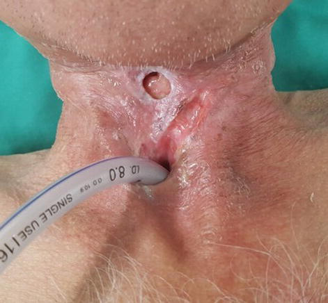
Obstruction
generated by esophageal protrusion of the voice prosthesis (See picture bellow)
Voice prosthesis' hood (Vega) protruding into the narrow esophagus.
The free flap that is sometimes used to replace the larynx has no peristalsis, making swallowing even more difficult. After surgery in such cases food descends to the stomach mostly by gravity. The time for the food to reach the stomach varies between individuals and ranges from 5 to 10 seconds.
Chewing the food well and mixing it with liquid in the mouth prior to swallowing is helpful, as is swallowing only small amounts of food each time and waiting for it to go down. Drinking liquids between solid foods is helpful in flushing down the food. Other strategies that can improve swallowing include head rotation, effort full swallow, siting
fully upright while eating/drinking and staying upright for at least 30-45
minutes afterwards, alternating food and liquid to help flush and clear foods
through the neopharynx, and swallow multiple times for each bite of food.
Eating takes longer; one must learn to be patient and take all the time needed to finish the meal.
The swelling that develops after surgery tends to decrease over time, which reduces the narrowing of the esophagus and ultimately makes swallowing easier. This is good to remember because there is alwayshope that swallowing will improve within the first few months after surgery. However, if this does not occur dilatation of the esophagus is one of the therapeutic option. Dilatation is usually done by an otolaryngologist or gastroenterologist (see below in the Dilation of the esophagus section).
Temporary placement of nonmetal expandable stents can be effective for
managing refractory benign strictures. If the problem persists pharyngeal
reconstruction may be needed. This can be accomplished by obtaining a flap of
non-radiated tissue (i.e., forearm) to create a wider throat.
Obstruction
generated by protrusion of the voice prosthesis into the esophagus can be
alleviated by replacing the prosthesis to one without a hood; making sure that
the prosthesis’ length is correct and that it is not “pistoning”, and/or by
changing the location of the puncture to a new site.
Swallowing difficulties can have a significant psychological impact on
laryngectomees, leading to anxiety, frustration, and social isolation. Fear of
choking or aspiration can also affect their eating habits and quality of life.
Management and treatment of swallowing difficulties
Management and treatment of swallowing difficulties in laryngectomees
often involve a multidisciplinary approach, including speech therapists,
otolaryngologists (ear, nose, and throat specialists), and dietitians.
Some strategies to help manage swallowing difficulties may include:
Swallowing Therapy: Speech therapists can provide exercises and techniques to improve
swallowing function and coordination of swallowing muscles.
Modified Diet: Laryngectomees may need to follow a modified diet consisting of soft or
pureed foods and thickened liquids.
Postural Techniques: Certain postural adjustments during eating and drinking, such as chin
tucks or head tilts, can help facilitate swallowing.
Saliva Substitutes: Artificial saliva or saliva stimulants may be recommended to alleviate
dry mouth and improve oral lubrication during swallowing.
Behavioral Modifications: Eating slowly, taking smaller bites, and thoroughly chewing food can
help laryngectomees manage swallowing difficulties.
It's essential for laryngectomees to work closely with healthcare
professionals to develop an individualized management plan tailored to their
specific needs and challenges. Regular follow-up appointments and adjustments
to treatment strategies may be necessary to optimize swallowing function and
improve quality of life.
Tests used for the evaluation of swallowing difficulty
Swallow tests are commonly used in
laryngectomees to evaluate and manage swallowing difficulties, which are a
frequent complication after total laryngectomy. There are five major tests that
can be used for the evaluation of swallowing difficulties:
Videofluoroscopy
Fiberoptic Endoscopic Evaluation of
Swallowing (FEES)
The specific test is chosen according to
the clinical condition.
Videofluoroscopy (VF) which is usually
the first test done to most patients, records swallowing during fluoroscopy.
(Picture 38) It allows accurate visualization and study of the sequence of
events which make up a swallow; it is limited to the cervical esophagus. The
video, taken from both the front and the side, can be viewed at a slower speeds
to enable accurate study. This helps identify abnormal movement of food, such
as aspiration, residue, strictures, pooling, and movement of anatomic
structures, muscle activities, pharyngeal constrictor spasm, pharyngoesophageal
stricture, and exact oral and pharyngeal transit times. It can also evaluate
the voice prosthesis status. The effects of various barium consistencies and
positions can be tested. Thick or solid food boluses can be used for patients
who complain of solid food dysphagia.
Indications for Swallow Testing
Swallow tests are typically performed in
laryngectomees when they report or exhibit signs of dysphagia, such as:
Difficulty swallowing solids or liquids
Coughing or choking during meals
Regurgitation or nasal regurgitation
Prolonged meal times
Weight loss or dehydration
Additionally, swallow tests may be
conducted routinely after total laryngectomy, even in asymptomatic patients, to
assess swallowing function and identify any potential issues early.
Timing of Swallow Tests
The timing of swallow tests in
laryngectomees can vary based on institutional protocols and individual patient
factors. Some common time points include:
Preoperatively: To establish a baseline
swallowing function
1-3 weeks postoperatively: To evaluate
for early complications like fistulas or strictures
Follow-up evaluations: At regular
intervals (e.g., every 6-12 months) or if new swallowing symptoms develop
By performing swallow tests, healthcare
professionals can accurately diagnose swallowing disorders in laryngectomees,
guide appropriate interventions (e.g., dietary modifications, swallowing
therapy, dilation), and monitor the effectiveness of treatment over time.
How to remove (or swallow) food stuck in the throat or the esophagus
Some laryngectomees experience recurrent episodes of food becoming stuck in the back of their throat or esophagus and preventing them from swallowing. Clearing the stuck food can be accomplished using these methods:
1. First do not panic. Remember that you cannot suffocate because as a laryngectomee, your esophagus is completely separate from your trachea.
2. Try to drink some liquid (preferably warm) and attempt to force the food down by increasing the pressure in your mouth. Sometimes changing the position of the head while swallowing to the right or left or bending the neck allows the stuck food to be move down into the stomach. If this does not work - 3.Use a finger to press on the middle and sides of your neck a little above or the sides of your stoma and try to swallow. Bend your head backwards while swallowing. If this does not work- 4.If you wear an HME hold it firmly with one hand and move it in a circular pattern and try to swallow. If this does not work-
5. If you speak through a tracheo-esophageal voice prosthesis try to speak forcefully. You can also brush and flush the prosthesis with water using flushing bulb. The air or water you blow. Try this first standing up and if it does not work bend over a sink and try to speak. If this does not work -
6. Bend forward (over a sink or hold a tissue or cup over your mouth), lowering your mouth below the chest and applying pressure over your abdomen with your hand. This forces the contents of the stomach upward and may clear the obstruction.
7. Swallowing a mashing a banana and drinking a sip of water may help to get something stuck in the throat down.
8. Try to plush the stuck food with water using feeding bolus syringe or 20-30 cc syringe connected to feeding tube or other catheter.
These methods work for most people. However, everyone is different and one needs to experiment and find the methods that work best for them. Swallowing does, however, get better in many laryngectomees over time.
Some laryngectomees report success in removing the obstruction by gently massaging their throat, walking for a few minutes, jumping up on their feet, sitting and standing several times, hitting their chest or the back, using a suction machine with the catheter paced in the back of their throat, or just waiting for a while until the food is able to descend into the stomach on its own.
Individuals whose tongue was partially or completely removed often experience swallowing difficulties. Applying pressure with the fingers bellow the chin and then sliding the hand down until the food is released may work. Enclosed is a video that demonstrates this method:
If nothing works and the food is still stuck in the back of the throat it may be necessary to be seen by an otolaryngologist or go to an emergency room to have the obstruction removed.
How to swallow and avoid food from being stuck in the esophagus or
throat
Swallowing as a laryngectomee requires patience and care. Following an episode of obstruction in the upper esophagus obstruction swallowing may be difficult for a day or two. This is because of local swelling in the back of the throat; normally, this will disappear with time.
The issues that contribute to swallowing difficulties are discussed in the Swallowing Difficulties section (see below).
Ways to avoid such episodes:
Eating slowly and patiently and avoid distractions
Taking small bites of food and chewing very well before swallowing
Swallowing a small amount of food at a time and always mixing it with liquid in the mouth before swallowing. Warm liquid makes it easier to swallow.
Attempting to swallow in a different position of the head such as bending the head down. This may work.
Moisten dry/crumbly foods with sauces, gravies, olive oil, margarine, or butter.
Some food items (soft diet items) are easier to swallow (i.e., soup and stews, yogurt, gelatin made with soft canned fruit, cooked vegetables, cottage cheese, scrambled eggs, macaroni and cheese, mashed potatoes with gravy, blended food, ice cream, custard, pudding, cooked cereal without lumps, banana, etc.).·
Soften hard foods such as toast by dunking it into milk, cocoa, coffee or tea.Chop, puree or blend foods that are difficult chewing or swallowing.
Flushing the food with more liquids as needed. (Warm liquids may work better for some individuals in flushing down the food)
Siting upright while eating/drinking, and stay upright for at least 30-45 minutes after mealtime to avoid reflux.
Avoiding food that ishard to chew or that can be stuck (i.e. peanuts).
Base of tongue strengthening exercises can help with generating more power to the swallow.
One needs to find out for him/her self what food is easier to ingest. Some foods are easy to swallow (e.g., toasted or dry bread, yogurt, and bananas) and others tend to be sticky ( e.g.,unpeeled apples,lettuce and other leafy vegetables, and steak).
Swallowing tablets and capsules
Ingestion of large pills and capsules may be difficult for laryngectomees. Over time, laryngectomees generally learn the maximal size of pills and capsules that they can swallow.
Tips to take medications include:
Some medications are available in several dosages which may be manufactured in smaller size pills or capsules. It is therefore possible to swallow the desired dose by taking several small pills or capsules.
Drinking fluid before swallowing a pill or capsule and taking a single one at a time.
The size of pills of some generic medications may vary depending on their manufacturers. If this is the case it may be possible to find a smaller size pill produced by a different manufacturer.
Some medications are also available as a suspension. It is best to check with one’s physician and pharmacy if a commercially available suspension is available, or can be prepared by one's pharmacy.
Pills can be crushed and dissolved in room temperature liquids, or broken down to small pieces prior to ingestion. Ingestion of crushed tablets is best done with food to slow their absorption. Slow release medications may lose their time delay action when crushed. Ingestion of large gel capsules may not be possible.
Capsules can be opened and their content swallowed. However, this exposes some medication to the stomach acidity that may inactivate and reduce their potency.
Some crushed medications or capsule contents may be irritating to the mouth and/or esophagus and stomach. Take them with food and flush them with liquids.
It is best to check with one’s physician and/or pharmacist if dissolving a pill or capsule’s contents is an option.
When oral ingestion is not an option other routes of drug administration may be possible. These include intramuscular and intravenous injection, aerosols, and rectal or vaginal suppositories.
When the size of a tablet or capsule is too large the physician may select a similar medication (from the same class or with similar effects) that is available in smaller size pill or capsule.
Attempt to swallow in a different position of the head such as bending the head down. This may work.
Narrowing (strictures) of the neopharynx and esophagus and their treatment
A stricture of the esophagus is a narrowing along the pharyngo-esophagus that blocks or inhibits the ease of food passage, resulting in the esophagus having an hour-glass configuration. Strictures after laryngectomy can be due to the effects of radiation as well as the tightness of the surgical closure and can also occur gradually as scarring develops. Interventions that can help the patient include:
Alternatives to these procedures include nasal enteric tubes, gastrostomy tubes, and
jejunostomy tubes. Total parenteral nutrition can also be used in patients who
are not candidates for these therapeutic options.
Esophageal
stents
An esophageal stent is a flexible mesh tube,
approximately 2cm (3/4 inch) wide, and is placed through the constricted area
of the esophagus to allow food and beverages to pass from the mouth to the stomach.
The stent is not as wide or as flexible as a normal esophagus and care must be taken not to block it while eating.
Stent
placement usually requires both endoscopic and fluoroscopic guidance, but can
be done with either modality safely. In general, dilation of a stricture before
placing the stent is not required. Most stents are placed distally and across
the gastroesophageal junction, but proximal stent placement (which requires
more precise placement) can also be performed. Complications include bleeding
and perforation (which are rare) as well as migration, tumor overgrowth, and
tumor ingrowth (which are more common).
Esophageal stent in place
Dilation of the neopharynx andesophagus
Narrowing of the neopharynx (The surgically reconstructed new pharynx) and esophagus is a very common consequence of radiation treatment as well as laryngectomy; and dilatation of the esophagus is often needed to reopen it. The procedure usually needs to be repeated and the frequency of this procedurevaries among individuals. In some people this is a lifelong requirement and in others the neopharynx and esophagus may stay open after a few dilations. The procedure requires local or systemic sedation or anesthesia because it is painful. A series of dilators with greater diameter are introduced into the esophagus to dilate it slowly. While the process breaks down the fibrosis, the condition may return after a while. Swelling at the
site of the dilation may make swallowing more difficult for several days
following the procedure.
Sometimes a balloon rather than a long dilator is used to dilate a local stricture. Another method that may help is the use of topical and or injectable steroids to the esophagus. Although dilation is done by an otolaryngologist or a gastroenterologist, in some cases it can be accomplished by the patient at home by performing self-dilation at-home device. In difficult cases, surgery may be needed to remove the stricture or replace the narrow section with a graft (tissue flap).
Because dilation breaks down fibrosis, the paingenerated by the procedure may last for a while. Taking pain medication can ease the discomfort.
Dilation can
cause temporary difficulties lasting a few days in speaking to those using
esophageal or voice prosthesis speech. The
stretching of the esophagus can cause temporary swelling (edema) of the esophageal
walls creating secretions that can block the voice prosthesis. Cleaning the
prosthesis with a brush and flushing it can reopen it making speaking possible.
Because the
walls of the pharyngoesophagus must vibrate to produce tracheoesophageal
or esophageal voice, dilation can stretch the esophagus to the extent that the
walls no longer contact and the result is a weaker voice. It typically will improve
as the effects of the dilation wear off, but then swallowing may worsen.
Voicing problems when using tracheoesophageal
speech
Laryngectomees using tracheoesophageal speech can develop a strained, halted or aphonic voice. These difficulties can
be due to pharyngeal constrictor spasm, post-operative edema (swelling), inappropriate
occlusion of the stoma, voice prosthetic issues, and pharyngoesophageal stricture.
Pharyngeal constrictor spasm or
pharyngeal hypertonicity: Pharyngeal constrictor spasm occurs when
the patient attempts to voice and the pharynx responses by tightens to prevent
or limit airflow up through the vibratory segment. This can result in no sound
(complete closure of the pharynx with spasm); a brief sound followed by no
sound (immediate release of air followed by complete closure of the pharynx
with spasm); or a strained vocal quality (partial closure of the pharynx with
spasm). In almost all instances this is not something that develops over time
and a laryngectomee will either have this issue post-operatively or will not.
Post-operative edema: Swelling of the tracheoesophageal walls following surgery can
narrow and even close the vibratory segment of the pharyngoesophagus and cause
voicing issues similar to when pharyngeal constrictor spasm is present. Voicing
difficulties dissipate over time as the edema subsides.
Inappropriate occlusion of the stoma when
voicing: How a patient occludes the stoma for voicing
can alter the quality of his/her voice. Digital occlusion with an incomplete seal
will lead to a weaker voice quality with turbulent air escape further
compromising intelligibility. Too much pressure with digital occlusion can
compress the vibratory segment and cause hypertonicity or complete inability to
speak (aphonia). If excessive digital pressure during occlusion is the issue,
patient training of light touch for occlusion is all that is needed.
Voice prosthetic issues affecting
voicing: There are several factors related to the voice
prosthesis that can cause a voice quality that mimics that of pharyngeal
constrictor spasm. A prosthesis that is partially or completely clogged by
dried mucus can cause a strained voice or inability to speak. Removing the
mucus will eliminate the issue and enable normal voicing.
An inappropriately-fitted voice
prosthesis can also cause similar vocal difficulties. A voice prosthesis that
is too long may press against the esophageal wall reducing or preventing
airflow through the device. A voice prosthesis that is too short may result in
narrowing of the esophageal side of the tracheoesophageal tract causing a
strained voice or aphonia. Voice prosthetic issues can be ruled out by removing
the voice prosthesis, making sure the appropriate sized dilator can pass easily
through the tracheoesophageal tract and testing the vocal quality once the
dilator is removed through an open tract. If the voicing difficulty persists
with open tract voicing prosthetic issues have been ruled out.
Swallowing difficulties as a cause of
voicing problems: Difficulty or inability to swallow
solids, may be due to blockage of the swallowing tube caused by a stricture. Dilatation
of the neopharynx and esophagus (see above) can improve phonation. However,
this may require repeated dilations as narrowing may recur.
Fluoroscopy is used to diagnose both
pharyngeal constrictor spasm (see below) and pharyngoesophageal stricture when speaking and
swallowing. Pharyngeal constrictor spasm appears as a transient bulge in the
posterior pharyngeal constrictor muscles when attempting to speak. Stricture, however,
is identified during swallowing and appears as a narrowing of the neopharynx or
esophageal lumen as contrast material transits through them. These two
conditions can also co-occur.
Identifying the exact underlying issue
causing the laryngectomee’s voice disturbances and appropriate intervention can
allow the patient to achieve fluent tracheoesophageal voicing.
Use of Botox® for pharyngeal
constrictor muscle spasm
Botox® is a pharmaceutical preparation of toxin A
which is produced byClostridium botulinum, an
anaerobic bacteria that causes botulism, a muscle paralysis illness. The
botulinum toxin causes partial paralysis of muscles by acting on their
presynaptic cholinergic nerve fibers through the prevention of the release of
acetylcholine at the neuromuscular junction. In small quantities it can be used to temporarily paralyze muscles for 3-4 months. It is used to control muscle spasms, excessive blinking, and for cosmetic treatment of wrinkles. Infrequent side effects are generalized muscle weakness and rarely even death. Botox®
injection is the treatment of choice for selected laryngectomees to improve
swallowing and tracheo-esophageal speech.
Indications for Botox® in Laryngectomees are:
Disturbed relaxation of the pharyngoesophageal
segment, causing difficulty with tracheoesophageal puncture (TEP) speech
production.
Pharyngoesophageal spasm leading to dysphagia
(swallowing difficulties).
Pharyngoesophageal spasm causing central valve
leak
Dysphonia (voice/speech problems) due to
pharyngoesophageal hypertonicity.
Benefits of Botox® Injection are:
Improves relaxation of the pharyngoesophageal
segment, facilitating better airflow for speech production.
Reduces air pressure required for speech,
making it easier to produce tracheoesophageal speech.
Increases maximum phonation time (ability to
sustain vowel sounds for longer).
Improves swallowing function by reducing
pharyngoesophageal spasm.
Reduces central valve leak around the voice
prosthesis.
A pharyngeal
constrictor muscle hypertonicity or pharyngoesophageal spasm (PES) is a common
cause for crico-pharyngeal dysfunction leading to tracheo-esophageal speech
failure after laryngectomy. In almost
all cases, this develops immediately after laryngectomy and not over time.
Constrictor muscle hypertonicity can increase peak intra-esophageal pressure
during speaking, thus interfering with fluent speech. It may also disturb
swallowing by interfering with the pharyngeal transit of food and liquids
Botox® can
reduce the hypertonicity and spasm of the lower jaw and vibrating segment muscles.
Botox is only effective for overactive muscles and may require the injection of
relatively large doses into the spastic muscles. It cannot help conditions that
are not due to muscle spasms, such as esophageal diverticula, strictures due to
fibrosis after radiation, and scars and narrowing after surgery.
Botox® injection can be carried out by otolaryngologists in the clinic. The injection can be done percutaneously (using electromyography),or through an esophago-gastro-duodeno-scope. The percutaneous injection into the pharyngeal constrictor muscles along one side of the newly formed pharynx (neopharynx) is done just above and to the side of the stoma.
An injection through an esophago-gastro-duodeno-scope can be performed whenever a percutaneous injection is not feasible. This method is used in patients with severe post-radiation fibrosis, disruption of the cervical anatomy, and anxiety or inability to withstand a percutaneous injection. This method allows direct visualization and greater precision. The injection into the PES segment is often done by a gastroenterologist and is followed by gentle expansion by balloon massage to facilitate uniform distribution of the Botox®.
Botox® injection
Pharyngo-cutaneous fistula
A pharyngo-cutaneous fistula is an abnormal connection between the pharyngeal mucosa to the skin. Typically a salivary leak develops from the pharyngeal area to the skin, indicating a breakdown of the pharyngeal surgical suture line. It is the most common complication after laryngectomy and usually occurs 7-10 days after the operation. The
main causes leading to development of fistula is poor wound healing. Risks factors leading to fistula are excessive tension
on the wound, surgical wound infection, poor nutrition, acid reflux or
gastroesophageal acid disease (GERD), tumor stage, type of surgery,
hypothyroidism, diabetes, continued use of alcohol and tobacco, and recent
radiation or chemotherapy therapy. .
Most fistulae
will heal on their own without additional surgery. The initial care is conservative and includes no
oral feeding through a naso - gastric tube
or percutaneous endoscopic gastrostomy); antibiotics; and wound care. Other
methods include sterilizing the fistula from within with 0,25% acetic acid by
mouth and wound care with negative-pressure,
hydrocolloid, hydrogel or silver coated dressings. Hyperbaric oxygen (HBO) may be used when conventional treatment fails.
Surgical
repair consisted of direct suture of pharyngo - cutaneous, closure with local,
pedicled or free flaps, and is reserved for fistulae that did not close on
their own and when the presence of the fistula pose some danger to the stoma
and the lungs.
The closure of the fistula can be evaluated by a dye test (such as ingestion of methylene blue which
appears in the skin if the fistula is unobstructed)and/or by radiographic contrast studies.
Pharyngo-cutaneous fistula
Smelling after laryngectomy
Laryngectomees may experience difficulties with their sense of smell. This may makes it hard to appreciate pleasant smells (i.e., food , flowers, perfumes), and can interfere with the ability to detect warning smells such as smoke or natural gas. Installing smoke and gas detectors is an essential precaution. There are numerous medications, diseases,
hormonal disturbances and chemicals that can disrupt the sense of smell,
sometimes permanently.
Smoke detector
Smelling difficulties occur despite the fact that regular laryngectomy surgery, does not involve nerves related to smell and the sense of smell or olfaction, remains intact. What has changed, however, is the pathway of airflow during respiration. Before a laryngectomy, air flows into the lungs through the nose and mouth. This movement of air through the nose allows for scents to be detected as they come in contact with the nerve endings in the nose responsible for the sense of smell.
After a laryngectomy, however, there is no longer an active airflow through the nose. This can be perceived as a loss of smell. Patients,
however, can relearn how to smell, by closing their mouths and swallowing to
create a vacuum that introduces air into the nose.
The “polite yawn technique” can also help laryngectomees regain their capacity to smell. This
method is known as the “polite yawn technique” because the movements involved
are similar to those used when one attempts to yawn with a closed mouth. Swift, downward movement of the lower jaw and tongue, while keeping the lips closed, will create a subtle vacuum, drawing air into the nasal passages and enabling the detection of any scent through the new airflow. With practice, it is possible to achieve the same vacuum using more subtle (but effective) tongue movements.
Video of the “polite yawn technique”
How
to avoid unpleasant smells
One
“advantage” of being a laryngectomee is the ability to avoid unpleasant smells or
odors such as cigarette smoke. This is because all our air intake is through the
stoma, while smelling is done through our nostrils. This can be achieved by
preventing the noxious smell from reaching the nose by:
Squeezing
the nostrils with the fingers
Placing
a cotton ball in each nostril
Wearing
a surgical mask over the nose with or without a fitted piece of plastic
Wearing a surgical mask
Squeezing the nostrils
Basic skills for laryngectomees by Elizabeth Finchem
Here are a few basic skills our newest laryngectomees can work on at home. They include a few topics that may help to discover how you can compensate for what some may think they have lost after laryngectomy. Recently a laryngectomee stated that he still can’t smell, taste, blow, or talk hands free prompted me to write about how to correct this perception with basic information.
There was a time, not so long ago, when the International Association of Laryngectomees Annual Meeting program included ‘speech improvement’ breakout sessions that provided tiered training for designated groups (beginner, intermediate, or advanced speakers) to improve their skills for whichever method they chose to communicate with year after year. Some folks referred to these sessions as “Jim’s Dog and Pony Show” since Jim Shanks, PhD, SLP, taught all of them at one point or another. Along the way the demonstrations included how learning to smell, taste and blow related to speech techniques we could use to improve the intelligibility of our speech after laryngectomy. I also learned the finer points of fluent esophageal speech while attending these demonstrations that amounted to very practical hands on labs for learning and teaching. I hope sharing some of these tried and true techniques will be helpful for your rehabilitation, and for those who are learning how to work with laryngectomees.
A good place to begin is to have you put your lips together and puff out your cheeks with only the air that is in your mouth. (voice prosthesis users do not need to inhale or exhale lung air to do this task.) Additional air will build in the mouth from your nasal passages. Notice how you can move the air from one cheek to the other. Next, you can use this air in your mouth to blow out the match. No, you don’t need to blow with lung air from your stoma for this task either. As you blow out the match you may find that you can smell the sulfur and the smoke if you sniff. Next you can try blowing out a candle in the same way. This mouth air can also be directed into a balloon one mouthful at a time to fully inflate a big balloon. You may discover that you can do things you’d assumed were no longer possible post op.
Following the above directions probably surprised you. They proved that you do have plenty of air in your mouth when you put your lips together. You have access to more air from your nose, which is still open and connected to your mouth. You have enough mouth air to blow out a match or candle. With your lips together drop your lower jaw. With these two steps you have created a bellows effect that draws air up your nasal passages so you can smell on demand as you sniff. It may be faint at first, but you can develop this skill to increase your ability to smell. This will also enhance you sense of taste since smell plays an important part of taste sensation. When this skill is developed some horn players have discovered how to use this ‘circular breathing (nose to mouth) to play their beloved horns again, using lung air for breathing only. Did you know that DIDGERIDOO players use this technique to play their tree trunk horns continuously? Check it out on youtube.com
It seems to me that very few SLPs, or speech instructors teach these basic steps anymore. As I begin working with new students this material seems to be very new to them. In fact, some seem to assume these skills are now impossible post op. I can tell you from my decades of teaching new laryngectomees as well as SLPs who wish to learn how to work with laryngectomees these basic methods do work. It makes me sad to read that so many are left to believe that their ability to taste, smell, or blow from the mouth or nose was taken along with their larynx. We can compensate and learn to do these maneuvers in a new way. They may not be as strong on the first few attempts, but they can be developed and serve the purpose very well.
You may wonder how you can possibly sniff up your nose or blow your nose again? Try it. Take those first few steps. Lips together and drop you lower jaw to draw air in with a sniff action, or reverse it by bringing your lower jaw back up as you use your tongue to thrust the air upward through the nose to blow one side at a time.
It is a pity when there are those who are left to believe that everything we did pre-operatively is gone forever. Slowly some discover what they are able to do with what is left on their own. Even so, few understand how it is possible, or how the body’s systems actually work. Another case of “go home and figure it out”? I hope not since we can learn from one another when we share information.
The
value of esophageal speech and how to master it.
by
Elizabeth Finchem
Esophageal
Speech (ES) has the following advantages:
1. A natural, biological source that
remains post-laryngectomy.
2. It is fully functional when mastered.
3. Requires no puncture or
tracheal-esophageal prosthesis.
4. Offers much less danger of aspiration
than TE puncture voice restoration, but not electro larynx..
5. It has pitch and volume control; though
at times somewhat limited.
6. Can offer speech on demand when one is
proficient.
7. Eliminates emergency voice loss, or a
need for voice prosthesis changes, or electrolarynx repairs.
Let
us be clear about what ES is and is not. For example, it is not gulping air and
burping up a word. That is the way I was
taught at the beginning of my speech therapy, and it sent me down the wrong
track. This method begins with too much
air and quickly leads to a cricopharyngeus sphincter (sphincter used when
swallowing) spasm. The only benefit from the burping concept is the feeling of
the air pressure rising and timing it to say a word or two with that air. What is more important in that action is the
slight tightening of the diaphragm that keeps the air moving “north” up into
the esophagus instead of forcing it into the stomach. It is esophageal speech, not stomach speech.
I
teach ES with a different approach.
After decades of training, developing my own ES voice, and experience
teaching others how to achieve ES in about 8-10 week. We begin with the basic
three methods of air intake:
1)
Consonant injection,
2)
Tongue press,
3)
Inhalation.
Inhalation
is not inhaling a breath into the lungs, but instead it is merely the opening
the cricopharyngeal sphincter (which is normally contracted). This is what you
do when the physician asks you to say “ah”.
You open this sphincter enough for examination of the mouth. You should hear a little “click” sound as the
wet tissue opens, and then you may have taken in enough air to say “ah”. Note that the tongue is always up in the
mouth for “tongue press”, and the tongue is always down in the mouth for
“inhalation”.
Instead
of focusing on reading word lists aloud that begin with consonants to inject
air into the esophagus we begin by focusing on double vowel that follows the
consonant sounds to release the air charge with Pee, Tee and Kay.
These three consonants coupled with vowels like ie (as in Pie), ea (as
inteach), or oo (as in took) seems
to be a better balance of air in, sound out.
After a few attempts it is important to take a sip or two of water to
keep the swallowing sphincter relaxed enough to let the consonant air back up
without a huge burp and rush of words …only to be “out of air”. Then what?
We
begin again with “P” to illustrate
that “Puh” (as in pup) sound can be
felt on the fingers when held out away from the mouth about 6 inches. According to Isaac Newton, “for every action
there is an opposite and equal reaction”.
The same amount of air felt on the fingers is also going backward into
the esophagus for the word “Pie”. Next comes the consonant T to teach the tongue placement against the front teeth at the gum
line. “Tie” should work here.
Working
with “K” (as in “Kay”) is important because it is on the
back of the tongue and may introduce too much air at first. For several reasons K, hard C and G may need
to be softened or modified a bit for the right balance of air injected.
Note
that it’s not unusual for a laryngectomee who has been over-articulating while
mouthing words with hope of being heard along with lip reading, or an electro
larynx user, to strike K, hard C (as in cake) and G (as in goat) harder than
they need to for ES. Only the consonants
are heard unless an occasional vowel is released inadvertently. More often there is a good deal of stoma
blast that goes with the mouthing in a failed attempt to be louder.
As
soon as we can move on to multiple syllables we begin working on phrases that
can be used daily. Rather than
“practice” word lists, we focus on using ES for simple questions and responses.
Pet commands such as sit, stay, come, down along with the pet’s name is
something that will be used often each day.
Family names and a few other phrases such as “thank you”, “car keys”,
and “get gas” can be tailored for “homework” that will be used. (For
conversation the electro larynx is best in the beginning when one is looking to
acquire and master it.) The interrogatives (who, what, when, where, why and
how) can keep a chat going while one uses their ES. We want these simple responses to become
spontaneous. In closing it is important
to be aware that expecting people to read lips is not helpful to learning ES. The goal is to learn how to produce the vowel
sounds that are now missing due to the loss of the normal sound generator: the
voice box. It is possible to learn to
produce esophageal vowel sounds by injecting enough air into the esophagus to
cause vibration that will become the new esophageal sound generator. People are always surprised to learn that
proficient ES speakers do not breathe when they speak. Instead they have learned to inhale, exhale,
inject and then speak. Otherwise we hear a good deal of stoma blast as they try
to use lung air to speak as they did before laryngectomy that is, while
exhaling. The stoma blast sound may
actually mask their ES vowel sounds as well as their consonants that
distinguish one word from the next. We
aim for intelligibility as well as effortless speech.
ES
is unimaginable for some who believe there must be something that has to
replace the larynx (voice box) that was removed. The truth is the human body is adaptable
enough to use what is left in a new way very effectively. Some consider this
second natural voice a miracle. I
do. Now I am sharing this gift with
others as many others shared this knowledge with me.


No comments:
Post a Comment